External Beam Radiation Therapy Treatments
The goal of radiation therapy is to get enough radiation into the body to kill the cancer cells while preventing damage to healthy tissue. There are several ways to do this. Depending on the location, size and type of cancer, you may receive one or a combination of techniques. Your treatment team will help you to decide which treatments are best for you.
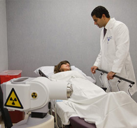
Radiation therapy can be delivered in two ways, externally and internally. During external beam radiation therapy, the radiation oncology team uses a machine to direct high-energy X-rays at the cancer. Internal radiation therapy, or brachytherapy, involves placing radioactive sources (for example, radioactive seeds) inside your body.
External Beam Radiation Therapy
During external beam radiation therapy, a beam of radiation is directed through the skin to the cancer and the immediate surrounding area in order to destroy the main tumor and any nearby cancer cells. To minimize side effects, the treatments are typically given five days a week, Monday through Friday, for a number of weeks. This allows doctors to get enough radiation into the body to kill the cancer while giving healthy cells time each day to recover.
The radiation beam is usually generated by a machine called a linear accelerator. The linear accelerator, or linac, is capable of producing high-energy X-rays and electrons for the treatment of your cancer. Using high-tech treatment planning software, your treatment team controls the size and shape of the beam, as well as how it is directed at your body, to effectively treat your tumor while sparing the surrounding normal tissue.
Several special types of external beam therapy are discussed in the next sections. These are used for specific types of cancer, and your radiation oncologist will recommend one of these treatments if he or she believes it will help you.
Three-Dimensional Conformal Radiation Therapy (3D-CRT)
Tumors are not regular — they come in different shapes and sizes. Three-dimensional conformal radiation therapy, or 3D-CRT, uses computers and special imaging techniques to show the size, shape and location of the tumor. Computer assisted tomography (CT or CAT scans), magnetic resonance imaging (MR or MRI scans) and/or positron emission tomography (PET scans) are used to create detailed, three-dimensional representations of the tumor and surrounding organs. Your radiation oncologist can then precisely tailor the radiation beams to the size and shape of your tumor with multileaf collimators (see picture, right) or custom fabricated field shaping blocks. Because the radiation beams are very precisely directed, nearby normal tissue receives less radiation and is able to heal quickly.
Intensity Modulated Radiation Therapy (IMRT)
Intensity modulated radiation therapy, or IMRT, is a specialized form of 3D-CRT that allows radiation to be more exactly shaped to fit the tumor. With IMRT, the radiation beam can be broken up into many “beamlets” and the intensity of each beamlet can be adjusted individually. Using IMRT, it may be possible to further limit the amount of radiation that is received by healthy tissue near the tumor. In some situations, this may also allow a higher dose of radiation to be delivered to the tumor, potentially increasing the chance of a cure.
Stereotactic Radiotherapy
Stereotactic radiotherapy is a technique that allows your radiation oncologist to precisely focus beams of radiation to destroy certain types of tumors. Since the beam is so precise, your radiation oncologist may be able to spare more healthy tissue. This additional precision is achieved by using a very secure immobilization, such as a head frame used in the treatment of brain tumors. Stereotactic radiotherapy is frequently given in a single dose (sometimes called radiosurgery) although certain situations may require more than one dose. In addition to treating some cancers, radiosurgery can also be used to treat malformations in the brain’s blood vessels and certain noncancerous (benign) neurologic conditions.
Sometimes a high dose of stereotactic radiotherapy can be focused upon a tumor outside the brain and given in a few treatments (typically three to eight). This form of treatment is called stereotactic body radiation therapy.
Image Guided Radiation Therapy (IGRT)
Radiation oncologists use image guided radiation therapy, or IGRT, to help better deliver the radiation to the cancer since tumors can move between treatments due to differences in organ filling or movements while breathing. IGRT involves conformal radiation treatment guided by imaging, such as CT, ultrasound or X-rays, taken in the treatment room just before the patient is given the radiation treatment. All patients first undergo a CT scan as part of the planning process. The imaging information from the CT scan is then transmitted to a computer in the treatment room to allow doctors to compare the earlier image with the images taken just before treatment. During IGRT, doctors compare these images to see if the treatment needs to be adjusted. This allows doctors to better target the cancer while avoiding nearby healthy tissue. In some cases, doctors will implant a tiny marker in or near the tumor to pinpoint it for IGRT.